

Pain intervention
Radiofrequency Techniques
Professor Dr. Ricardo Ruiz-López is a pioneer in the development and application of advanced Radiofrequency techniques for the treatment of painful pathologies of the spine and peripheral nervous system, developing and perfecting techniques that are now part of the routine clinical practice in numerous specialized centers.
As founder and former president of the World Institute of Pain, Dr. Ruiz-López has played a fundamental role in training specialists and in the global dissemination of the highest standards of excellence in interventional pain management.
Conventional and pulsed radiofrequency techniques are minimally invasive procedures used to treat chronic pain by targeting specific nerves with radiofrequency energy. Both are performed under radiological guidance and allow for precise, safe, and outpatient treatment. The choice between conventional or pulsed radiofrequency is made on an individual basis after a thorough clinical evaluation, selecting the most appropriate technique according to the origin and characteristics of each patient’s pain. These treatments are frequently combined with regenerative medicine techniques.
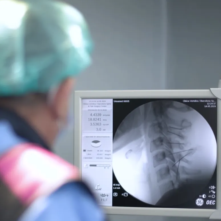
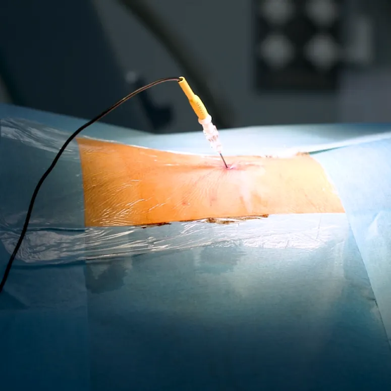
Conventional Radiofrequency (Thermal)
Main indications :
- Herniated discs or pain of discogenic origin
- Lower back and neck pain of facet joint origin.
- Vertebral osteoarthritis.
- Sacroiliac pain.
- Specific neuralgias.
- Chronic pain in certain joints.
Advantages :
- Prolonged relief of symptoms
- Reduction in the consumption of analgesic medication.
- Fast recovery without the need for complex open surgery.

Pulsed Radiofrequency
Main indications :
- Sciatica and radiculopathies.
- Occipital neuralgia.
- Neuropathic pain syndrome.
- Persistent postsurgical pain.
Advantages :
- It does not produce significant thermal damage to the nerve.
- Especially useful in neuropathic pain.
- It can be applied to nerves where it is desired to fully preserve sensory and motor function.

RF OF THE GASSERIAN GANGLION (TRIGEMINAL)
Trigeminal neuralgia is one of the most intense painful conditions that can occur. It manifests as episodes of sudden, very intense facial pain, usually described as an electric shock or a stabbing sensation, which can last from a few seconds to several minutes. In some cases, the pain can become continuous and persist for much of the day.
Painful attacks can be triggered by everyday stimuli as simple as talking, chewing, brushing teeth, or shaving. Due to the intensity and frequency of the episodes, this condition can cause significant functional limitations and a marked decline in quality of life.
When drug treatment does not provide adequate symptom control or loses effectiveness over time, minimally invasive techniques can be used. Radiofrequency thermocoagulation of the Gasserian ganglion is currently the most effective and safe procedure for treating trigeminal neuralgia.
This technique involves inserting a specialized cannula through the cheek, guided by radiological imaging, to the Gasserian ganglion, a structure located at the base of the skull that is part of the trigeminal nerve. Once the correct position is reached, radiofrequency energy is applied in a controlled manner to very precise areas of the ganglion, interrupting the transmission of the signals responsible for pain.
The procedure is performed in an operating room under sedation, on an outpatient basis and without the need for hospital admission. Thanks to its high success rate and low risk of complications, it is one of the leading therapeutic options for patients with trigeminal neuralgia that is refractory to pharmacological treatment.

Pulsed RF of the glossopharyngeal nerve
Glossopharyngeal neuralgia is a rare neurological condition characterized by intense pain in areas innervated by the glossopharyngeal nerve, primarily the throat, tonsillar region, pharynx, and occasionally the ear. This pain typically presents as acute and recurrent episodes that can significantly impact a patient’s quality of life.
When conventional drug treatments fail to provide adequate symptom relief, minimally invasive interventional techniques may be considered. Among these, pulsed radiofrequency ablation of the glossopharyngeal nerve is an effective and safe alternative for pain management.
The procedure involves inserting a thin cannula, guided by imaging techniques, into the vicinity of the glossopharyngeal nerve through a small puncture in the lateral region of the neck, behind the ear. Once its correct placement is confirmed, pulsed radiofrequency energy is applied at low temperature for several minutes, modulating the transmission of pain signals without causing significant nerve damage.
It is an outpatient, minimally invasive and well-tolerated technique, whose effects usually begin to be appreciated progressively in the days following treatment, providing significant pain relief in a large number of patients.

Pulsed RF of the sphenopalatine ganglion
Radiofrequency ablation of the sphenopalatine ganglion is a minimally invasive technique used to treat certain types of facial pain and difficult-to-control headaches. It is particularly indicated for patients with facial neuralgia and some types of primary headache, such as cluster headache (also known as Horton’s headache), when conventional treatments have not provided satisfactory relief.
It can also be beneficial in other headaches of vascular origin, such as migraine, as well as in chronic paroxysmal headache, especially in those cases refractory to pharmacological treatment.
By applying controlled radiofrequency to the sphenopalatine ganglion, it is possible to modulate the transmission of the signals involved in the generation and maintenance of pain, helping to reduce the intensity and frequency of attacks and improving the quality of life of patients.
Pulsed RF for painful scars or post-surgical neuropathic pain
In some patients, pain may persist after surgery or appear later in the operated area. In many cases, this pain has a neuropathic origin, resulting from damage or irritation of nerve structures, and can be particularly difficult to treat.
Among the most frequent examples are persistent pain after inguinal hernia surgery, plastic surgery procedures, or orthopedic procedures. It can also occur after fractures that have left lasting damage or caused tissue adhesions. In these situations, further surgery does not always resolve the problem and, sometimes, can even worsen symptoms by creating new scars or increasing nerve irritation.
Pulsed radiofrequency is a minimally invasive therapeutic alternative that has proven to offer satisfactory results in numerous patients with chronic neuropathic pain. The procedure is performed under sedation and consists of the precise insertion of one or more cannulas near the affected nerves, without the need for open surgery.
By applying pulsed radiofrequency energy, the transmission of pain signals is modulated, reducing pain intensity and improving the patient’s functionality. The effects usually begin to be noticeable in the days following treatment and can last for months or, in certain cases, provide long-term, lasting relief.
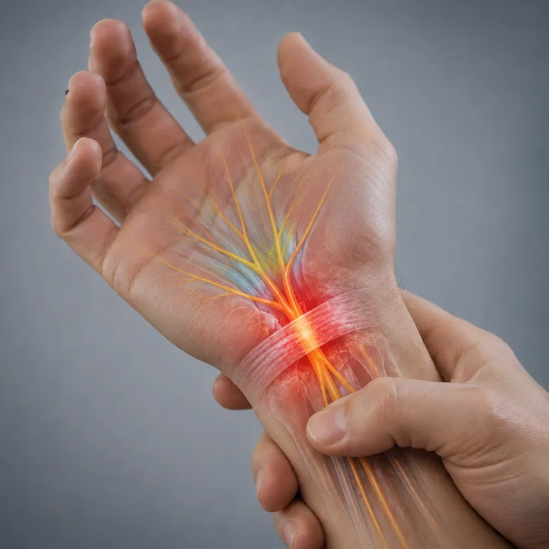
Pulsed RF of the median nerve or carpal tunnel syndrome (Ruiz-López technique)
Carpal tunnel syndrome, caused by compression of the median nerve at the wrist, can now be treated with minimally invasive techniques, avoiding the need for conventional surgery in many cases.
At Clínica Vertebra we perform the treatment using Dual Pulsed Radiofrequency, an advanced and safe technique that allows modulation of the median nerve activity without causing nerve damage or requiring surgical incisions.
This procedure offers excellent clinical results in selected patients, with significant improvement in pain, tingling, and numbness, maintaining its benefits long-term. Furthermore, it is a repeatable technique, requiring no subsequent rehabilitation and with a very rapid recovery time, typically allowing a return to daily activities within 24–48 hours.
The treatment is performed in an operating room under sedation, by inserting two thin radiofrequency cannulas carefully positioned next to the median nerve in the wrist. Pulsed radiofrequency is then applied precisely and in a controlled manner through these cannulas.
This technique was initially described by Dr. Ricardo Ruiz-López, a pioneer in the development of interventional pain treatments, and its results have been published in leading international scientific journals.
RF lumbar or cervical sympathectomy for complex regional pain syndrome (CRPS) or reflex sympathetic dystrophy
Complex Regional Pain Syndrome (CRPS), also known internationally as Complex Regional Pain Syndrome, is a chronic pain condition that usually affects one limb, generally following trauma, fracture, surgery, or nerve injury.
This is a complex pathology of the nervous system in which there is an alteration in the mechanisms of pain processing and regulation, involving both the peripheral and central and autonomic nervous systems. When the origin is specifically related to a demonstrable nerve injury, the condition may be called causalgia.
Patients experience intense, persistent pain that is disproportionate to the initial injury, frequently accompanied by sensory disturbances, inflammation, changes in skin temperature and color, excessive sweating, joint stiffness, and progressive functional limitations. Trophic changes may also occur in the skin, hair, and nails. In many cases, the pain extends beyond the initially affected area.
The functional and emotional impact of CRPS is often very significant, potentially affecting quality of life and being associated with anxiety, sleep disturbances, or depressive symptoms.
The diagnosis is primarily clinical and requires a specialized assessment of pain and neurological and vascular function in the affected limb, using a comprehensive, personalized, and minimally invasive approach. To this end, VERTEBRA SpinePain & Regenerative SURGERY CENTERS has high-resolution, highly specific diagnostic equipment, including an integrated Computerized Infrared Telethermography System for obtaining pathognomonic images of dystrophic disease (CRPS I).
The course of CRPS is variable and depends largely on early diagnosis and treatment. Some patients experience progressive recovery, while in others the pain can become chronic and lead to significant functional limitations if not treated appropriately.
Treatment should be approached in a multidisciplinary and individualized manner, combining specific rehabilitation, advanced physiotherapy, pharmacological treatment and interventional pain techniques.
When conservative treatments fail to adequately control symptoms, advanced techniques on the sympathetic nervous system, such as Radiofrequency Sympathectomy, can be used.
This procedure involves selectively modulating or interrupting nerve conduction in the sympathetic chain responsible for maintaining pain. Treatment is performed at the cervical level in cases affecting the upper extremities, or at the lumbar level when the pain affects the lower extremities.
Before definitive treatment, a diagnostic block of the sympathetic chain is usually performed using local anesthetics, with the aim of confirming the sympathetic origin of the pain and assessing the therapeutic response.
The procedures are performed in an operating room under radiological control and sedation, using minimally invasive and outpatient techniques, allowing the patient to return home a few hours after treatment.
In selected patients and refractory cases, other advanced therapies may also be considered, such as:
- Spinal cord stimulation
- Advanced Neuromodulation
- Implantable intrathecal drug infusion systems
Facet denervation by RF of medial branches of the cervical spine (rhizolysis)
Cervical rhizolysis or cervical facet denervation by radiofrequency of medial branches is a minimally invasive technique indicated for the treatment of chronic cervical pain originating in the facet joints of the cervical spine.
The facet joints, also called zygapophyseal joints, are small joints located at the back of the cervical vertebrae. These structures can degenerate or become inflamed over time due to processes such as cervical osteoarthritis, mechanical overload, trauma, or degenerative changes, becoming a significant source of neck pain and cervicobrachialgia.
Radiofrequency ablation allows for the selective targeting of the small nerve branches responsible for transmitting pain from these joints, through the controlled application of thermal energy. The goal of the treatment is to modulate or interrupt pain conduction, while preserving normal motor function and sensation in the upper extremities.
This is a highly precise and safe procedure, performed under radiological control, in which only specific sensory nerve fibers involved in the transmission of pain are treated.
The procedure is performed in an operating room on an outpatient basis. The patient receives intravenous sedation to ensure maximum comfort during the procedure.
Using high-precision radiological guidance, the cervical levels responsible for the pain are identified. Local anesthesia is then administered, and thin, specialized radiofrequency cannulas are inserted to reach the selected facet nerve branches. Radiofrequency energy is then applied in a controlled manner through these cannulas to perform neuromodulation or therapeutic thermocoagulation.
After the procedure, the patient remains under observation for a short period of time and can usually return home approximately one hour after the treatment.
It is common to notice mild discomfort or a feeling of local inflammation in the puncture area during the first few days, which is generally controllable with conventional analgesic treatment.
The main advantages of cervical rhizolysis include:
- minimally invasive technique
- Outpatient procedure
- Rapid recovery
- Absence of open surgery
- Preservation of cervical mobility
At VERTEBRA SpinePain & Regenerative SURGERY CENTERS we perform advanced spinal radiofrequency treatments using image-guided precision techniques, with a specialized focus on the interventional treatment of cervical and spinal pain.
Facet denervation by RF of medial branches of the dorsal spine (rhizolysis)
Dorsal rhizolysis or dorsal facet denervation by radiofrequency of medial branches is a minimally invasive technique indicated for the treatment of chronic dorsal pain originating in the facet joints of the thoracic spine.
The facet joints, also known as zygapophyseal joints, are small joints located at the back of the spine that allow for movement and stability between the vertebrae. Degenerative processes such as osteoarthritis, facet joint inflammation, postural abnormalities, or certain mechanical overloads can turn these joints into a significant source of persistent back pain.
Radiofrequency allows for precise targeting of the small nerve branches responsible for transmitting pain from these joints. Through the controlled application of thermal energy, selective neuromodulation or thermocoagulation of the sensory fibers involved in pain transmission is performed, preserving motor function and normal sensation at all times.
This is a safe and highly precise procedure, performed under radiological control, which allows the specific treatment of the structures responsible for pain without the need for open surgery.
The procedure is performed in an operating room on an outpatient basis. Before the procedure, the patient receives intravenous sedation to ensure maximum comfort and safety.
Using high-precision radiological guidance, the vertebral levels responsible for the pain are identified. Local anesthesia is then administered, and thin, specialized radiofrequency cannulas are inserted to reach the selected facet nerve branches. Radiofrequency energy is then applied in a controlled manner through these cannulas to deliver the therapeutic treatment.
After the procedure, the patient remains under observation for a short period of time and can usually return home approximately one hour after the treatment.
It is common to notice mild discomfort or a feeling of inflammation in the treated area during the first few days, which is usually controllable with conventional analgesic medication.
The main advantages of dorsal rhizolysis include:
- minimally invasive technique
- Outpatient procedure
- Rapid recovery
- Absence of open surgery
- Preservation of spinal mobility
- Prolonged improvement of back pain in selected patients
At VERTEBRA SpinePain & Regenerative SURGERY CENTERS we perform advanced image-guided spinal radiofrequency techniques, offering personalized and minimally invasive treatments for chronic spinal pain.
Facet denervation by RF of medial branches of the lumbar spine (rhizolysis)
Lumbar rhizolysis or lumbar facet denervation by radiofrequency of medial branches is a minimally invasive technique indicated for the treatment of chronic lower back pain originating in the facet joints of the lumbar spine.
Lumbar facet joint pain is one of the most frequent causes of chronic lower back pain and is usually related to degenerative processes such as facet joint osteoarthritis, mechanical overload, vertebral instability or degenerative changes in the spine.
The facet joints, also called zygapophyseal joints, are small joints located at the back of the lumbar vertebrae that contribute to the stability and mobility of the spine. These structures are innervated by small sensory nerve branches responsible for transmitting pain sensations.
Radiofrequency ablation allows for the selective targeting of these nerve branches through the controlled application of thermal energy. The goal of the treatment is to modulate or interrupt the transmission of pain from the facet joints, while fully preserving motor function and normal sensation in the lower extremities.
This is a precise, safe and effective technique, performed under radiological control, which allows the specific treatment of the origin of the pain without the need for open surgery.
The procedure is performed in an operating room on an outpatient basis. Before the procedure, the patient receives intravenous sedation to ensure maximum comfort and safety during the treatment.
Using high-precision radiological guidance, the lumbar levels responsible for the pain are identified. Local anesthesia is then administered, and thin, specialized radiofrequency cannulas are inserted to reach the selected facet nerve branches. Radiofrequency energy is then applied in a controlled manner through these cannulas to perform neuromodulation or therapeutic thermocoagulation.
After the procedure, the patient remains under observation for a short period of time and can usually return home approximately one hour after the treatment.
During the first few days, a slight discomfort or inflammatory sensation may appear in the treated area, generally controllable with conventional analgesic medication.
The main advantages of lumbar rhizolysis include:
- minimally invasive technique
- Outpatient procedure
- Rapid recovery
- Absence of open surgery
- Preservation of lumbar mobility
- Prolonged improvement of lower back pain in selected patients
At VERTEBRA SpinePain & Regenerative SURGERY CENTERS we perform advanced image-guided spinal radiofrequency treatments, offering personalized and minimally invasive solutions for the treatment of lower back pain and degenerative spinal conditions.
Facet denervation by RF of medial sacroiliac branches (rhizolysis)
Radiofrequency rhizolysis of the sacroiliac joints is a minimally invasive technique indicated for the treatment of chronic pain originating in the sacroiliac joints.
Dysfunction or inflammation of the sacroiliac joints is a common cause of lower back pain, buttock pain, and even radiating pain to the pelvis or lower extremities. This type of pain can arise as a result of degenerative processes, mechanical overload, trauma, postural abnormalities, previous lumbar surgery, or biomechanical changes in the spine and pelvis.
The sacroiliac joints connect the spine to the pelvis and play a vital role in load transmission and lumbopelvic stability. These structures are innervated by small sensory nerve branches, responsible for transmitting pain sensations.
Radiofrequency ablation allows for selective targeting of these nerve branches through the controlled application of thermal energy. The goal of the treatment is to modulate or interrupt the transmission of pain from the sacroiliac joint, while fully preserving motor function and normal sensation in the lower extremities.
This is a precise, safe and effective technique, performed under radiological control, which allows the specific treatment of the origin of the pain without the need for open surgery.
The procedure is performed in an operating room on an outpatient basis. Before the procedure, the patient receives intravenous sedation to ensure maximum comfort and safety during the treatment.
Using high-precision radiological guidance, the anatomical areas responsible for transmitting pain from the sacroiliac joint are identified. Local anesthesia is then administered, and thin, specialized radiofrequency cannulas are inserted to reach the selected sensory nerve branches. Radiofrequency energy is then applied in a controlled manner through these cannulas to perform neuromodulation or therapeutic thermocoagulation.
After the procedure, the patient remains under observation for a short period of time and can usually return home approximately one hour after the treatment.
During the first few days, a slight discomfort or inflammatory sensation may appear in the treated area, generally controllable with conventional analgesic medication.
The main advantages of sacroiliac radiofrequency include:
- minimally invasive technique
- Outpatient procedure
- Rapid recovery
- Absence of open surgery
- Preservation of lumbopelvic mobility and stability
- Prolonged pain improvement in selected patients
At VERTEBRA SpinePain & Regenerative SURGERY CENTERS we perform advanced image-guided radiofrequency treatments for the management of sacroiliac pain and degenerative pathologies of the spine and pelvis, offering a personalized, precise and minimally invasive approach.
Pulsed RF of the greater trochanter
Radiofrequency ablation of the greater trochanter is a minimally invasive technique indicated for the treatment of chronic pain located in the lateral region of the hip, especially in patients with trochanteric pain syndrome, gluteal tendinopathy, or trochanteric bursitis refractory to conservative treatments.
Trochanteric pain is a common cause of hip pain and functional limitations, and can make everyday activities such as walking, climbing stairs, standing, or sleeping on the affected side difficult. In many cases, this pain is related to inflammatory or degenerative processes of the gluteal tendons and periarticular structures of the greater trochanter.
When conventional treatment through physiotherapy, medication, injections or rehabilitation does not achieve sufficient improvement, radiofrequency can offer an effective and minimally invasive therapeutic alternative.
Radiofrequency ablation works by applying controlled thermal energy to small sensory nerve branches responsible for transmitting pain from the trochanteric region. The goal of the treatment is to selectively modulate or interrupt pain transmission, while fully preserving muscle function and hip mobility.
The procedure is performed precisely under ultrasound and/or radiological control, allowing the anatomical structures responsible for the pain to be located and only the nerve fibers involved to be treated.
The procedure is performed in an operating room or interventional procedure room, on an outpatient basis and usually under mild sedation and local anesthesia to ensure maximum patient comfort.
Using image guidance, thin, specialized radiofrequency cannulas are inserted into selected areas around the greater trochanter. Radiofrequency energy is then applied in a controlled manner through these cannulas to perform therapeutic neuromodulation.
The procedure usually lasts a short time and the patient can return home a few hours after the treatment.
Recovery is usually quick, allowing you to gradually resume your normal activities within a few days. You may experience mild, temporary discomfort in the treated area for the first 24–72 hours.
The main advantages of radiofrequency ablation of the greater trochanter include:
- minimally invasive technique
- Outpatient procedure
- Rapid recovery
- Absence of open surgery
- Preservation of hip mobility
- Significant reduction in pain in selected patients
- Possibility of combining with regenerative therapies and functional rehabilitation
At VERTEBRA SpinePain & Regenerative SURGERY CENTERS we perform advanced image-guided interventional treatments for the treatment of hip pain and musculoskeletal pathologies, offering a personalized, precise and minimally invasive approach.
Pulsed RF of the suprascapular nerve (shoulder)
Pulsed Radiofrequency of the Suprascapular Nerve is a minimally invasive technique indicated for the treatment of chronic shoulder pain, especially in patients with degenerative, inflammatory or tendinous pathology who have not responded adequately to conservative treatments.
The suprascapular nerve plays a crucial role in transmitting pain signals from a large portion of the shoulder joint. Various conditions, such as rotator cuff tendinopathies, tendon ruptures, subacromial bursitis, glenohumeral osteoarthritis, adhesive capsulitis, or persistent pain following surgery, can cause intense pain and significant functional limitations.
When treatment through physiotherapy, rehabilitation, medication or injections does not achieve sufficient improvement, pulsed radiofrequency of the suprascapular nerve can offer an effective and safe therapeutic alternative.
Pulsed radiofrequency is an advanced neuromodulation modality that applies controlled pulses of electrical energy to the nerve without causing significant thermal damage. The goal is to modify the transmission of pain signals and reduce nerve sensitization, preserving the nerve’s functional integrity.
Unlike conventional thermal radiofrequency, pulsed radiofrequency works by using controlled electric fields and low temperatures, allowing pain to be treated without destroying the nerve or altering the motor function of the shoulder.
The procedure is performed on an outpatient basis, under sedation and local anesthesia, to ensure maximum patient comfort.
Using high-precision ultrasound and/or radiological guidance, the suprascapular nerve is located in the posterior region of the shoulder. A thin, specialized radiofrequency cannula is then inserted until it reaches the vicinity of the nerve, and sensory and motor stimulation is performed to confirm accurate and reliable localization.
Next, pulsed radiofrequency is applied in a controlled manner for several minutes to perform therapeutic neuromodulation.
Recovery is rapid and usually allows a gradual return to normal activities within a few days. In some cases, mild, temporary discomfort may occur at the injection site during the first 24–48 hours.
The main advantages of pulsed radiofrequency ablation of the suprascapular nerve include:
- minimally invasive technique
- Outpatient procedure
- Absence of open surgery
- Preservation of shoulder motor function
- Rapid recovery
- Significant reduction in pain and functional improvement
- Possibility of combining with physiotherapy and regenerative therapies
At VERTEBRA SpinePain & Regenerative SURGERY CENTERS we perform advanced image-guided neuromodulation and radiofrequency techniques for the specialized treatment of shoulder pain and musculoskeletal pathologies, offering a personalized, precise and minimally invasive approach.
Three-channel RF knee
Tri-channel Radiofrequency Knee is a minimally invasive technique indicated for the treatment of chronic knee pain, especially in patients with osteoarthritis, persistent pain after surgery or prosthesis, and in those cases where conservative treatments have not achieved adequate improvement.
Chronic knee pain is a leading cause of functional limitations and reduced quality of life. Degenerative processes such as osteoarthritis can cause inflammation, joint wear, and persistent pain that limits everyday activities like walking, climbing stairs, or standing for extended periods.
Three-channel radiofrequency allows precise action on the sensory nerve branches responsible for transmitting pain from the knee joint, using an advanced image-guided neuromodulation technique.
The three-channel radiofrequency technique is based on the simultaneous and precise treatment of the main geniculate nerves responsible for the transmission of knee pain.
By applying controlled radiofrequency to these sensory nerves, it is possible to modulate or interrupt pain transmission, while preserving the stability, mobility, and motor function of the joint at all times.
The use of multichannel radiofrequency systems allows for more precise, homogeneous, and effective lesions, optimizing therapeutic coverage and improving clinical outcomes in selected patients.
The procedure is performed on an outpatient basis, usually under mild sedation and local anesthesia to ensure maximum patient comfort.
Using high-precision ultrasound and/or radiological guidance, the geniculate nerve branches responsible for pain transmission are identified. Subsequently, thin, specialized radiofrequency cannulas are inserted into the selected anatomical points.
Through the three-channel system, radiofrequency is applied simultaneously and in a controlled manner to several sensory nerves in the knee, performing therapeutic neuromodulation safely and precisely.
Recovery is usually quick, allowing you to gradually resume your normal activities within a few days. You may experience mild, temporary local discomfort in the treated area during the first 24–72 hours.
The main advantages of three-channel radiofrequency knee ablation include:
- minimally invasive technique
- Outpatient procedure
- Rapid recovery
- Absence of open surgery
- Preservation of joint mobility and stability
- Significant reduction in pain and functional improvement
- Therapeutic alternative in patients who are not candidates for surgery or prosthesis
- Possibility of combining with physiotherapy and regenerative therapies
At VERTEBRA SpinePain & Regenerative SURGERY CENTERS we perform advanced image-guided radiofrequency and neuromodulation treatments for the specialized treatment of joint and musculoskeletal pain, offering personalized, precise and minimally invasive solutions for chronic knee pain.
Pulsed radiofrequency of Morton's neuroma
Pulsed Radiofrequency for Morton’s Neuroma is a minimally invasive technique indicated for the treatment of chronic forefoot pain caused by irritation or entrapment of the interdigital nerves, known as Morton’s Neuroma.
Morton’s neuroma is a common foot condition that typically affects the nerves located between the toes, most often between the third and fourth interdigital spaces. This condition can cause intense pain, burning sensations, electric shock-like pain, tingling, or the feeling of “walking on a stone,” especially when walking or wearing tight shoes.
In many patients, pain can significantly limit physical activity and quality of life. When conservative treatment with orthotics, physical therapy, changes in footwear, medication, or injections does not achieve sufficient improvement, pulsed radiofrequency can offer an effective and safe therapeutic alternative without the need for surgery.
Pulsed radiofrequency is an advanced neuromodulation modality that applies controlled pulses of electrical energy to the affected nerve, without producing significant thermal destruction.
The goal of treatment is to modulate the transmission of pain signals and reduce nerve irritation responsible for the symptoms, preserving the structure and function of the nerve.
Unlike conventional surgical techniques, pulsed radiofrequency allows pain to be treated in a minimally invasive way, avoiding surgical scars and reducing recovery time.
The procedure is performed on an outpatient basis, usually under sedation and local anesthesia to ensure maximum patient comfort.
The affected interdigital nerve is located using high-precision radiological and/or ultrasound guidance. A thin, specialized radiofrequency cannula is then inserted to the vicinity of the neuroma, and sensory stimulation is performed to confirm its precise and safe location.
Next, pulsed radiofrequency is applied in a controlled manner for several minutes to perform therapeutic neuromodulation.
Recovery is usually quick, allowing you to gradually resume your normal activities within a few days. During the first 24–48 hours, you may experience mild, temporary discomfort in the treated area.
The main advantages of pulsed radiofrequency treatment for Morton’s neuroma include:
- minimally invasive technique
- Outpatient procedure
- Rapid recovery
- Absence of open surgery
- Preservation of nerve structure
- Significant reduction in pain and functional improvement
- Reduction of sick leave and recovery time
- Possibility of avoiding surgery in selected patients
At VERTEBRA SpinePain & Regenerative SURGERY CENTERS we perform advanced ultrasound-guided radiofrequency and neuromodulation treatments for the specialized treatment of neuropathic pain and foot pathologies, offering precise, personalized and minimally invasive solutions.
Pulsed radiofrequency of metacarpophalangeal joints
Pulsed Radiofrequency of the Metacarpophalangeal Joints is a minimally invasive technique indicated for the treatment of chronic pain in the hand and fingers, especially in patients with osteoarthritis, joint inflammation, post-traumatic pain or degenerative pathology that does not respond adequately to conservative treatments.
The metacarpophalangeal joints, located between the metacarpal bones and the proximal phalanges, play a fundamental role in the mobility, strength, and function of the hand. Various inflammatory and degenerative processes can cause persistent pain, stiffness, and functional limitations, hindering everyday activities such as writing, grasping objects, or performing fine motor skills.
When treatment with medication, rehabilitation, splints, injections or physiotherapy does not achieve sufficient relief, pulsed radiofrequency can offer an effective and safe therapeutic alternative.
Pulsed radiofrequency is an advanced neuromodulation modality that applies controlled pulses of electrical energy to the small sensory nerve branches responsible for transmitting joint pain, without producing significant thermal injury.
The goal of the treatment is to modulate the transmission of pain signals and decrease nerve sensitization, while fully preserving motor function and mobility of the hand and fingers.
Thanks to the use of high-precision ultrasound guidance, the treatment can be selectively performed on the structures responsible for the pain, minimizing tissue damage and improving the safety of the procedure.
The procedure is performed on an outpatient basis, usually under sedation and local anesthesia to ensure maximum patient comfort.
High-resolution ultrasound is used to identify the joints and nerve branches responsible for the pain. Then, thin, specialized radiofrequency cannulas are inserted into the selected areas, and sensory stimulation is performed to confirm the correct anatomical location.
Next, pulsed radiofrequency is applied in a controlled manner for several minutes to perform therapeutic neuromodulation.
The procedure is usually short and the patient can return home shortly after treatment.
Recovery is usually quick, allowing you to gradually resume your normal activities within a few days. You may experience mild, temporary discomfort in the treated area for the first 24–48 hours.
The main advantages of pulsed radiofrequency ablation of the metacarpophalangeal joints include:
- minimally invasive technique
- Outpatient procedure
- Rapid recovery
- Absence of open surgery
- Preservation of hand mobility and functionality
- Significant reduction in pain and functional improvement
- Possibility of combining with rehabilitation and regenerative therapies
At VERTEBRA SpinePain & Regenerative SURGERY CENTERS we perform advanced ultrasound-guided radiofrequency and neuromodulation treatments for the specialized treatment of joint and musculoskeletal pain, offering precise, personalized and minimally invasive solutions for hand and limb pathologies.
Contact us and regain your quality of life
