

Minimally invasive spine surgery
Percutaneous Lumbar Extraforaminoplasty (PLEF Technique Park/Ruiz-Lopez)
Percutaneous Lumbar Extraforaminoplasty (PLEF) is an advanced minimally invasive neurosurgical technique intended for the treatment of lumbar radiculopathy caused by foraminal and extraforaminal compression of the lumbar nerve roots.
This percutaneous technique, developed for the selective release of ligamentous and compressive structures of the lumbar foramen, represents an innovative therapeutic alternative in patients with radicular pain refractory to conventional pharmacological, rehabilitative and interventional treatments.
The PLEF Park/Ruiz-López technique is especially indicated in patients with degenerative lumbar foraminal stenosis, ligamentous hypertrophy, foraminal fibrosis and certain intraforaminal or mixed disc herniations, allowing the treatment of neural compression through a minimally invasive approach that preserves vertebral anatomy.

What is lumbar foraminal stenosis?
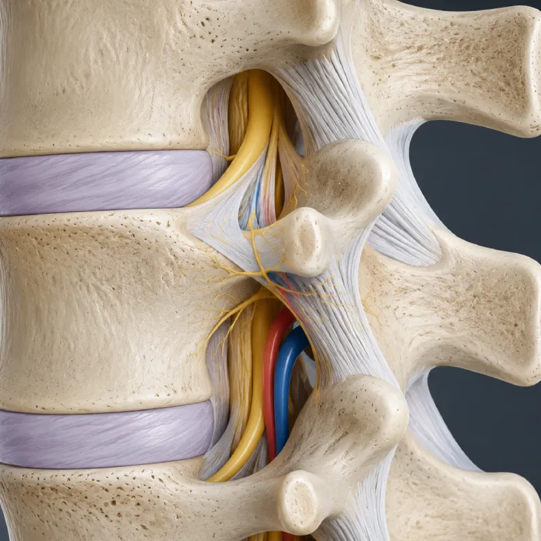
Lumbar foraminal stenosis consists of the progressive narrowing of the intervertebral foramen, that is, the exit canal of the lumbar nerve roots.
This narrowing can occur as a result of multiple degenerative processes, including:
- Decreased disc height
- Facet joint osteoarthritis
- Hypertrophy of the ligamentum flavum
- Fibrosis and foraminal adhesions
- Degenerative facet subluxation
- Foraminal or extraforaminal disc herniations
- Fibrous ring protrusion
Sustained compression of nerve roots can cause microvascular alterations and inflammatory phenomena responsible for lower back pain, sciatica, paresthesia, muscle weakness, neurogenic claudication and even progressive neurological deficit in the most advanced cases.
Basis of the PLEF technique
One of the fundamental pathophysiological concepts of this technique is the role of the lumbar foraminal ligaments and perineural adhesions in the genesis of radicular pain and chronic neural compression.
The lumbar foraminal ligaments—including the transforaminal and corporotransverse ligaments—participate in the stabilization and protection of nerve roots and vascular structures within the intervertebral foramen. However, degenerative changes, fibrosis, and abnormal adhesions can lead to nerve entrapment and vascular compromise of the nerve root.
The PLEF technique allows for selective percutaneous release of these compressive structures, promoting neural decompression and functional recovery of the affected root.
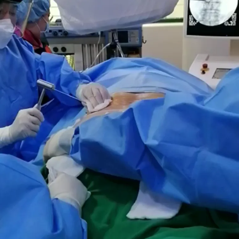
The procedure is performed in an operating room under high-precision radiological control and strict sterility measures.
The procedure is usually performed under intravenous sedation and local anesthesia, allowing a percutaneous approach through small millimeter incisions.
Through specialized transforaminal and extraforaminal access systems, the release of adhesions, fibrosis, and ligamentous structures responsible for neural compression is performed, preserving as much as possible the anatomical stability of the lumbar spine.
The technique can be combined, in selected cases, with disc decompression procedures, radiofrequency, ozone therapy or regenerative therapies.
Percutaneous lumbar extraforaminoplasty can be especially useful in patients with:
- Refractory chronic lumbar radiculopathy
- Degenerative lumbar foraminal stenosis
- Foraminal or extraforaminal disc herniations
- Neurogenic claudication
- Fibrosis and periradicular adhesions
- Failed back surgery syndrome
- Patients with borderline or alternative indications to instrumented surgery
Thanks to its minimally invasive nature, functional recovery is usually significantly faster compared to conventional open surgery.

The main advantages of the PLEF technique include:
- Advanced percutaneous neurosurgical technique
- Absence of extensive open surgery
- Preservation of vertebral stability
- Reduced muscle and tissue damage
- Faster functional recovery
- Reduction of radicular and lumbar pain
- Shorter hospital stay
- Possibility of avoiding or delaying instrumented surgery in selected patients
At VERTEBRA SpinePain & Regenerative SURGERY CENTERS we perform advanced percutaneous surgery techniques, neural decompression and image-guided spinal intervention, offering highly specialized and personalized treatments for complex degenerative pathologies of the spine.